The best neuroscience images from the Queensland Brain Institute for 2015 have been revealed.
Images were submitted into two categories: artistic and scientific.
The awards, judged by Dr Allison Holland from The University of Queensland’s School of Communication and Arts and renowned Brisbane artist Dr Svenja Kratz, highlight the point where art meets science in the laboratory.
Neuroscience images: art meets science
Dr Kratz said the scientific award went to Elise Roberts for her image titled 'Colour through the eye of a mantis shrimp', pictured above.
"The image best encapsulated and communicated the broader field of neuroscience and the research undertaken at the institute," she said.
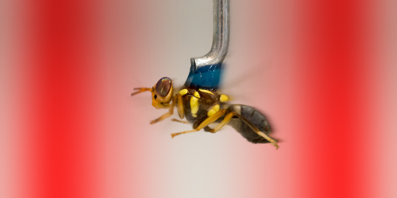
Kiaran Lawson, a PhD Student in the Srinivasan lab, won the artistic category, with his image of a fruit fly tethered to a metal rod flying swiftly through a virtual environment. The Srinivasan lab focusses on how vision in bees and flies use vision to shape their behaviour.

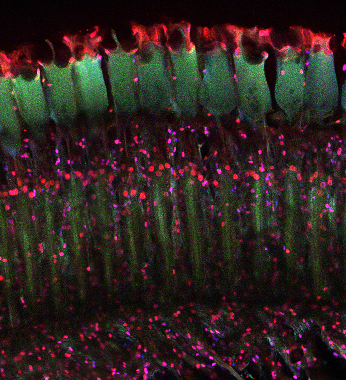
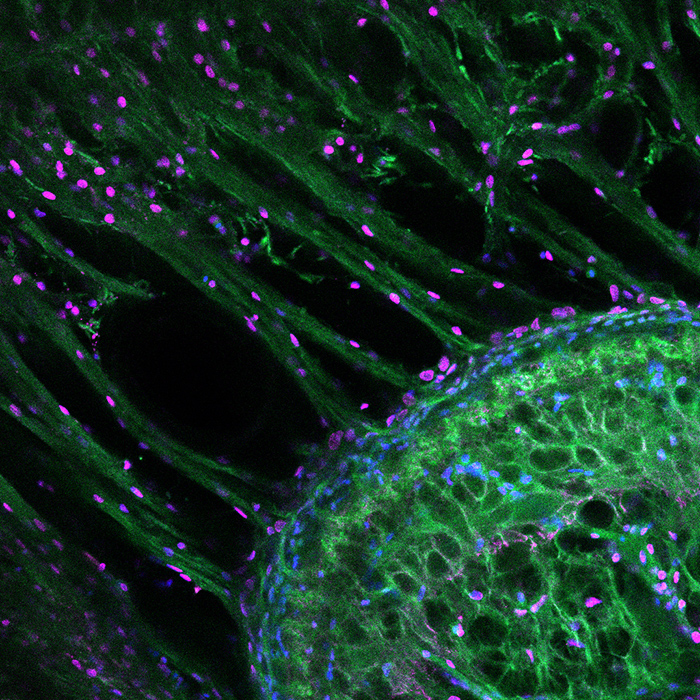
People's choice winner: A snapshot of developing dopaminergic neurons in embryonic rat midbrain. The neurons are dynamically marching from midline to their destinations down- and side-wards. Leon (Wei) Luan, Post-Doctoral Research Fellow, Eyles lab.
This is an image slice of the retina and neural tissue of a mantis shrimp enhanced through incubation with fluorescent antibodies. Research into these aggressive crustaceans is helping to pave the way for new biologically inspired technologies in areas like rapid cancer detection. (Credit:Trent Brooks-Richards, Honours Student, Marshall Lab)
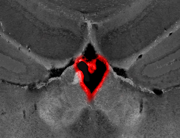
This is the fluid-filled ventricle in the middle of the brain, where a lining of cells (the choroid plexus) has come away, forming a heart shape. (Credit: Dana Bradford, Visiting Research Scientist, Burne Lab)
A cross section of the visual units in the compound eye of the mantis shrimp stained with fluorescent antibodies. (Credit:Trent Brooks-Richards, Honours Student, Marshall Lab)
This is an image slice of the retina and neural tissue of a mantis shrimp enhanced through incubation with fluorescent antibodies. (Credit:Trent Brooks-Richards, Honours Student, Marshall Lab)
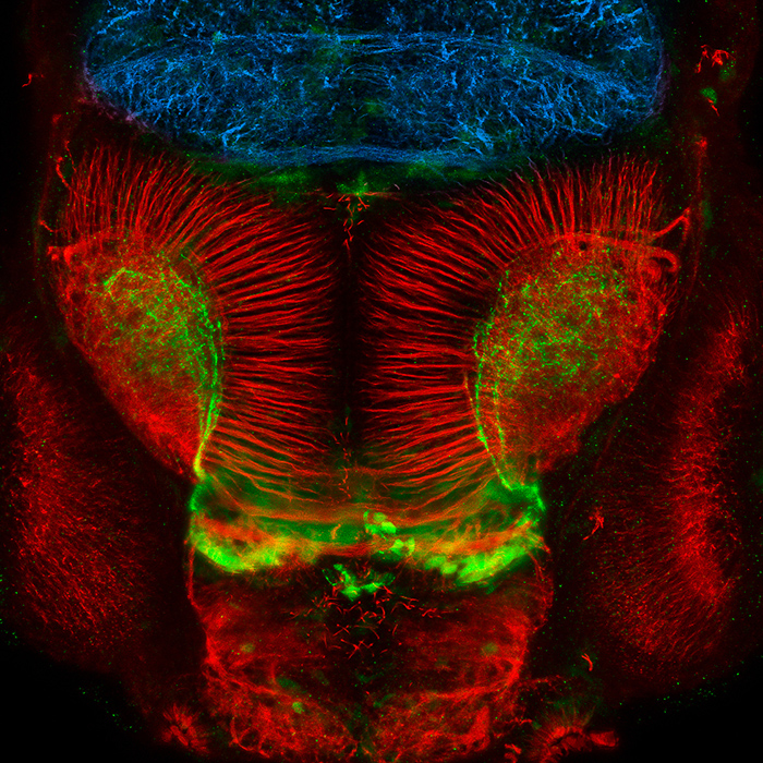
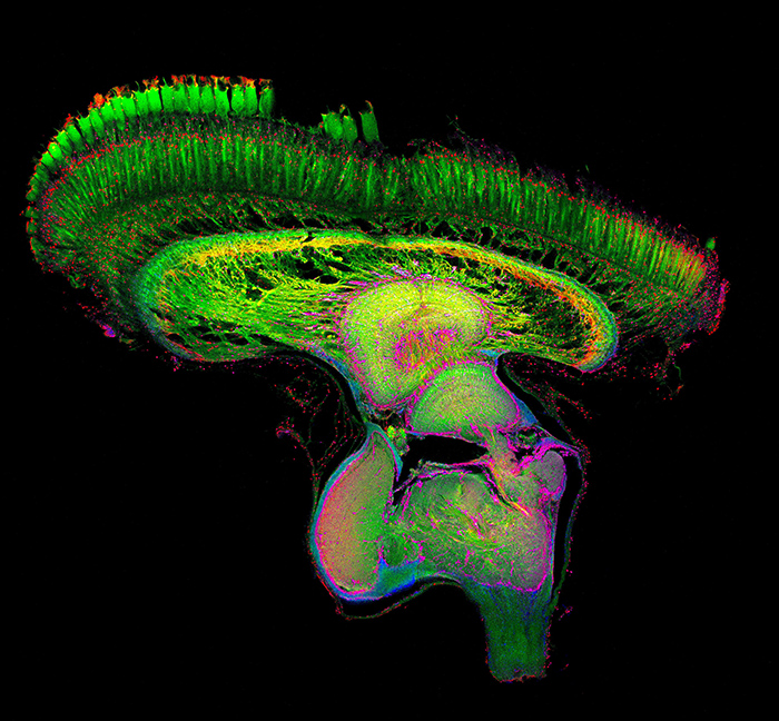
Representative image of the zebrafish brain at three days post-fertilization, including forebrain, optic tectum and midbrain. (Giacomotto Jean, Research Fellow, Mowry Lab)
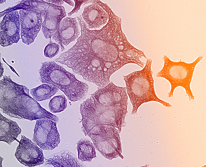
Neuroblastoma cells stained for tubulin reveal a complex cytoskeletal network. (Nadia Cummins, PhD student, Götz lab)
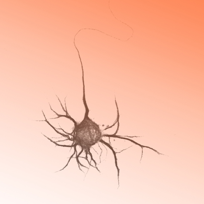
A young neuron, just starting to sprout its first processes. (Nadia Cummins, PhD student, Götz lab)
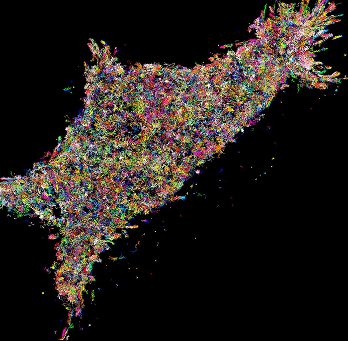
A super resolved photoactivated localisation microscopy image of single Munc18-1-mEos2 molecule trajectories in Munc18-1/2 double knockdown (DKD-PC12 cells). We have used a cutting edge technology of single particle tracking to study the dynamics of Munc18-1, Munc18-1Δ317-333, syntaxin-1A at a nanoscale level. (Ravikiran Kasula, PhD Student, Meunier lab)
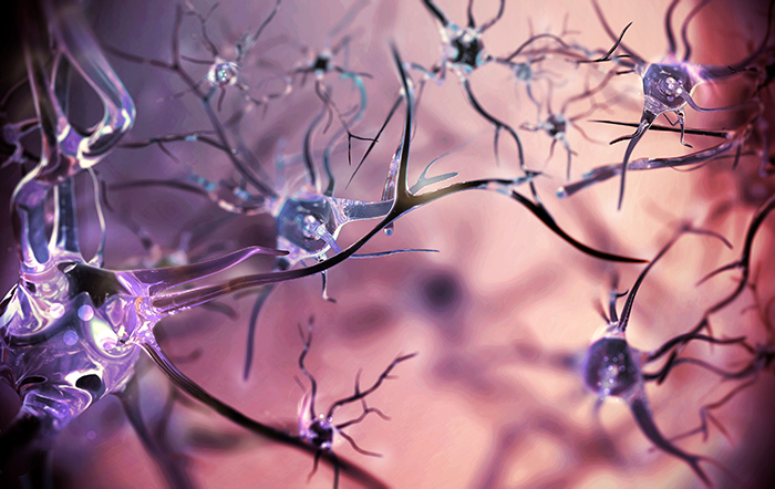
Representation of neuronal connectivity within the brain. (Anita Goldinger, PhD Student, Centre for Neurogenetics and Statistical Genomics)
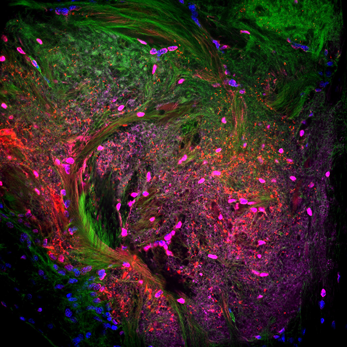
The optic lobes of the mantis shrimp, Lysiosquillina maculata. When incubated in fluorescent stains, this lobe reveals a colourful network of cell nuclei (blue), f-actin (green), synapsin (pink) and serotonin (red). (Credit: Elise Roberts, Honours Student, Marshall Lab).
MEDIA: QBI media and communications manager Bernadette Condren, b.condren@uq.edu.au, +61 7 3346 6353


